function eegplot(S,meas,sens,linestyle)
%EEGPLOT
plots a multi-channel data set
% eegplot(S,meas,sens,linestyle)
% S
is a matrix with m rows (channels) and n
coloms (timesamples)
% meas
is the montage used (optional)
% sens
is the y-axes scale (optional)
% linestyle
(optional)
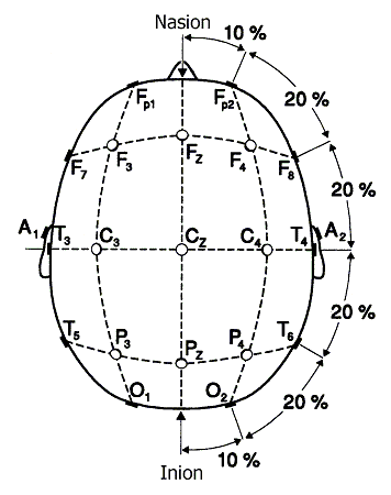
meas19chch: longitudinal montage used to review epileptic
events. This data structure can be fed into eegplot.
elpos19chch : A two-column matrix containing theta and phi
in radials. The standard electrode positions on a spherical head model
of the
19 electrodes. This information is necessary when performing EEG source
localization.
% |
|z
% |
|
% | |
th /
% |
|-->/
% |
|
/
% |
| /
y
% |
|/-----------
% |
/\
% |
/
\
% |
/`-->\
% |
/ phi
\
% |
/
% |
/x
The rows represent the
following electrodes:
'Fp1'
'Fp2'
'F3' 'F4'
'C3'
'C4' 'P3'
'P4'
'O1' 'O2'
'F7'
'F8' 'T3'
'T4'
'T5' 'T6'
'Fz'
'Cz' 'Pz'
Matlab code on ICA and
EEG source localization can be found on the open source package EEGLAB:
http://sccn.ucsd.edu/eeglab/
Raw data stored with
an average reference i.e. sum of all channels equals zero for each time
sample.
Sample frequency 256
Hz
spike1.mat : Is a 19 x 1281 matrix. The rows represent
the 19 channels (see above for order); the columns represent the time
samples.
>>eegplot(sig_av,meas19chch)
Two epileptic
transients can be observed, one at 1.6 s on channels T3-T5 and T5-O1,
both
peaks are pointing to each other, and one at 2.5 s at channels F8-T4
and T4-T6.
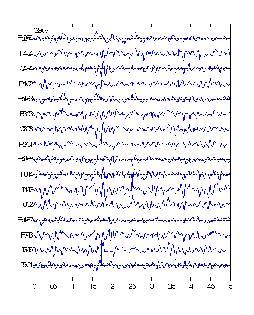
spike2.mat: Is a 19 x 1281 matrix as above.
>>eegplot(sig_av,meas19chch)

At 1.8 s an eye-blink
or eye-movement artifacts occurs mainly on channels Fp2-F4, Fp1-F3,
Fp2-F8 and
Fp1-F7. These are channels in the vicinity of the eyes. Epileptic
transients
are noticed at 2.5s and 4.4s on the channels Fp1-F7 and T5-O1. We also
observe
runs of high frequency activity mainly on channels T4-T6 and F8-T4.
This
signal may be due to muscle activity or
due to 50 Hz power line disturbance.
Ictal EEG
Matlab code:
function eegplotavr(S,meas,sens,linestyle)
%EEGPLOTAVR
plots a multi-channel data set in an
average reference montage
% eegplotavr(S,meas,sens,linestyle)
% S
is a matrix with m rows (channels) and n
coloms (timesamples)
% meas
is the montage used (optional)
% sens
is the y-axes scale (optional)
% linestyle
(optional)
meas21avr: average reference montage used to review
epileptic seizures. This data structure can be fed into eegplotavr.
sig_ictal.mat: Is a 21 x 2500 matrix. The rows represent the
21 channels; the columns represent the time samples. The sampling
frequency is
250 Hz.
>>eegplotavr(sig_ictal,meas21avr,100)
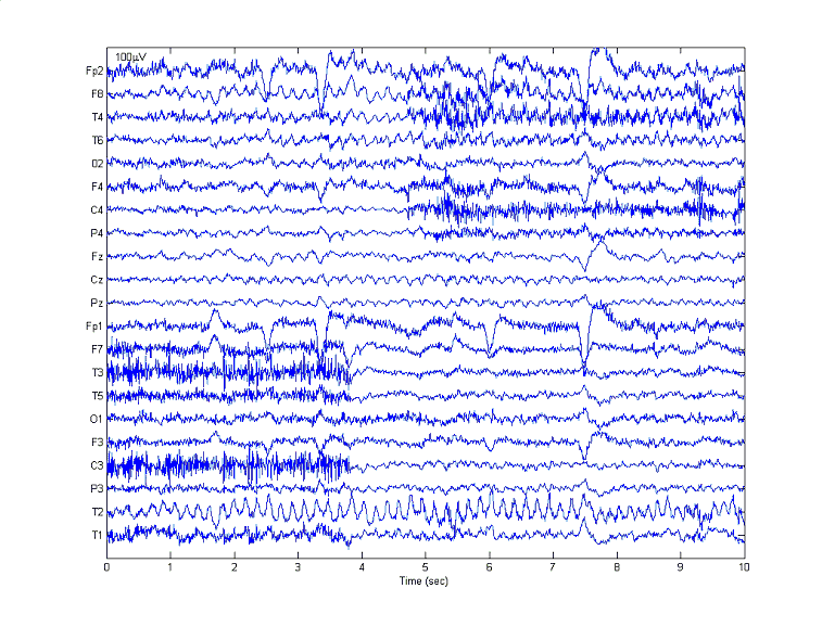
Epileptic seizure
activity can be observed on channels T2, F8, T4 and T6. At 2.5 s, 3.4
s, 6 s,
7.6 s eye blinks occur. Muscle artifacts can be observed between 0 s -
3.9 s on
channels F7, T3, T5, C3, T1 and between 5 s - 10 s on channels F8, T4,
F4, C4
and P4.